
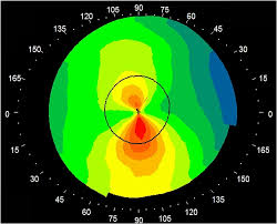
Topography is evaluated using Placido disk patterns or mires reflected off of the
tear film of the anterior cornea and converted to color scales. Because the image is
generated off the tear film, irregularities in tear film can significantly impact the
quality and fidelity of a Placido disk topography. Secondly, lack of patient fixation
may affect the quality of the topographic image. Finally, there is decreased
accuracy of posterior elevation values especially after refractive surgery.
Corneal Tomography
In comparison to topography which is good at capturing anterior corneal power,
overall corneal shape is best captured by tomography. Scanning slit technology,
Scheimpflug-based imaging, and anterior segment OCT technology is used to take
multiple slit images of the cornea and provide data on both the anterior and
posterior surfaces.
Uses
Corneal topography and tomography is most commonly used for the following
purposes
Refractive surgery: To screen candidates for normal corneal shape, patterns and
ruling out suspicious or keratoconic patterns . Post operatively , imaging can help
to assess the dioptric change created at corneal level ( thus the effective change in
the cornea) , ruling out de-centered or incomplete ablation , post excimer ectasia or
other changes.
Keratoconus : Early screening of keratoconus suspects is one of the most useful
roles of corneal imaging. Early keratoconus and suspects look normal on slit lamp
examination ,and the central keratometry (3 mm) gives only a limited assessment.
Therefore imaging has become the gold standard in screening keratoconus
suspects. In cases with established keratoconus, the role of topography and
tomography is paramount for monitoring progression and doing a timely collagen
cross linking , and in hard contact lens fittings.
Post surgery astigmatism : Post cataract surgery and post keratoplasty corneal
astigmatism can be studied with the topographer and selective suture removal or
other interventions can be planned.
Surgical planning in cases with corneal astigmatism : Limbal relaxing incisions
and other methods of topography guided incision placement are used by surgeons
to reduce post operative astigmatism.
Effect of corneal and ocular surface disorders: Disorders such as pterygium ,
limbal desmoids, corneal scars, or degenerations can cause changes in the corneal
curvature and irregular astigmatism.
Other uses : Contact lens fitting , incision placement and intrastromal ring
placement in keratoconus , monitoring of ocular vs corneal wavefront.
General Principles
Corneal imaging uses three of the following principles
Placido disc reflection
Scanning slit
Scheimpflug photography
Placido Disc Reflection for curvature analysis
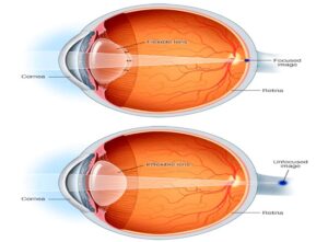
The primary optical aim of the cornea is refraction and focusing of the light rays as
it acts as a covering lens. However , all non-ideal refracting surfaces reflect some
light off them. This is the principle used for Purkinje imaging as well in Placido
discs. Placido disc is a device made of concentric rings drawn on a device of a
different color (generally white rings on a black background) . The first refracting
surface of the cornea (the tear film –air interface) acts as a convex mirror and
reflects back light in a pattern dependent of the corneal pattern. Topographers use
this technique to their advantage.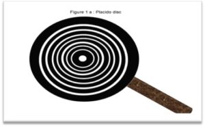 Whereas the original placido discs were aimed a
qualitative keratoscopy , the videokeratoscope or the topographer uses
mathematical formulae to provide a point-to-point quantitative gradient of these
subtle changes in topography.
Scanning Slit elevation evaluation
Scanning Slit is one of the elevation based methods for assessment of corneal
curvature and power. Multiple complimentary slits are used to perform an
assessment of the corneal surface. In the Orbscan , 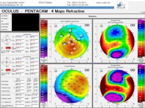 40 slits (20 each from nasal and
temporal side) are projected on the cornea to assess 240 points on each slit. The
triangulation between the reference slit beam surface and the reflected beam
captured by the camera can be used to analyze the anterior and posterior corneal
curvature and corneal thickness


