Introduction –
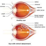
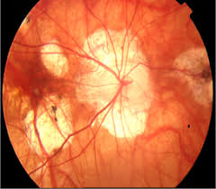
Myopia, commonly known as nearsightedness, has rapidly evolved from a standard refractive inconvenience into a global public health crisis. Epidemiological projections estimate that by 2050, nearly half of the world’s population—approximately 5 billion people—will be myopic, with up to 1 billion individuals at risk of high myopia. High myopia is not merely a requirement for thicker lenses; it significantly elevates the lifetime risk of sight-threatening ocular pathologies, including retinal detachment, myopic maculopathy, glaucoma, and early-onset cataracts. Historically, eye care practices treated myopia as a static optical defect, correcting clear central vision through single-vision concave lenses. However, groundbreaking vision science over the past three decades has shifted the focus from the center of the eye to its periphery. At the heart of this paradigm shift lies Relative Myopic Defocus (RMD)—a pivotal optical mechanism that regulates eye growth and forms the scientific bedrock of modern myopia control interventions.
1. The Basics of Refractive Errors and Ocular Growth
To understand relative myopic defocus, one must first grasp how the human eye regulates its physical growth—a process known as emmetropization. During early childhood, the human eye undergoes an active, homeostatic growth process designed to adjust its axial length to match the refractive power of the cornea and crystalline lens. The goal of this process is to ensure that parallel rays of light coming from distant objects focus precisely on the retina without any conscious accommodative effort.
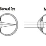
In an emmetropic (normally focusing) eye, incoming parallel light rays pass through the cornea and crystalline lens to converge precisely on the fovea, the center of the retina responsible for sharp, detailed visual acuity. When this balance is achieved, the individual enjoys crisp distance vision, and the sclera receives biological signals to maintain stable ocular dimensions.
Axial myopia primarily develops due to excessive axial length elongation—the eyeball grows too long from front to back along its anteroposterior axis. As the eye stretches physically beyond its ideal focal distance, light rays from distant objects converge in front of the retina rather than directly on it, creating a blurred central image. While genetic factors play a significant role in determining a child’s susceptibility to myopia, environmental triggers such as prolonged near-work activities, excessive screen time, and reduced exposure to natural outdoor light accelerate this physical elongation process.
2. Central Vision vs. Peripheral Refractive Error
For decades, optical science evaluated refractive error almost exclusively along the visual axis (the straight line passing through the pupil to the fovea). The implicit assumption was that central vision dictated ocular elongation and that as long as the fovea received a sharp image, the eye had no optical reason to grow unnaturally. However, seminal animal models and clinical research pioneered by researchers like Dr. Earl Smith III demonstrated a surprising truth: the peripheral retina plays a dominant role in guiding eye growth. Experiments involving primates revealed that even when the central fovea was optically ablated or masked, the eye continued to regulate its axial growth based purely on visual inputs received by the peripheral retina.
The central retina (fovea) occupies a very small anatomical region, spanning approximately 1.5 millimeters across, representing less than 5% of the total retinal area. Its primary function is high-resolution visual acuity, detailed color perception, and fine visual discrimination. In contrast, the peripheral retina covers over 90% of the total retinal surface area. While it possesses lower visual acuity, it is highly sensitive to motion, spatial luminance, and peripheral optical defocus. Because of its vast surface area, the peripheral retina generates the vast majority of visual feedback signals that drive scleral remodeling and ocular expansion.
The human eye is rarely a perfect sphere; it is often oblate or prolate in shape. Consequently, the peripheral retina often curves differently than the optical focal plane created by the eye’s cornea and lens. This fundamental difference creates two distinct peripheral optical states:
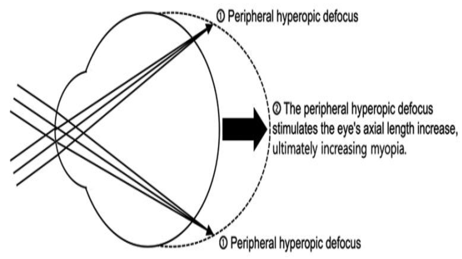
Peripheral Hyperopic Defocus: Incoming peripheral light rays focus behind the peripheral retina, creating a focal mismatch where the image plane lies posterior to the light-sensitive tissue.
Peripheral Myopic Defocus: Incoming peripheral light rays focus in front of the peripheral retina, creating an image plane that lies anterior to the light-sensitive tissue.
3. The Mechanics of Defocus: Hyperopic vs. Myopic
The eye uses visual defocus as an internal navigational map to determine whether it needs to grow larger or stop growing. The direction of this defocus dictates the biochemical response within the sclera (the outer white structural layer of the eye).
The Trap of Traditional Single-Vision Glasses
When an uncorrected myopic individual puts on standard single-vision spectacle lenses, the center of the image is brought sharply onto the fovea, restoring excellent central visual acuity. However, standard concave spherical lenses have a uniform negative power across their surface. Because the human retina curves forward in the periphery, these flat negative focal planes cause peripheral light rays to fall behind the peripheral retina.
This introduces artificial relative hyperopic defocus across the peripheral visual field. The peripheral retina interprets this focal plane behind itself as a signal that the eye is physically “too short” to meet its visual demands. In response, local retinal cells release chemical cascades that soften the sclera and promote tissue expansion, driving further axial elongation. Paradoxically, traditional single-vision glasses designed to correct vision ended up inadvertently signaling the eyeball to grow longer over time.
4. Biological Mechanisms: From Light to Chemical Signals
How does an optical image projected onto the retina alter the structural collagen matrix of the human eyeball? The emmetropization signal cascade operates primarily through local intraocular pathways, operating almost entirely independently of the central nervous system or optic nerve pathways.
Step 1: Photoreceptor Detection and Signal Processing
Amacrine cells, horizontal cells, and bipolar cells within the peripheral retinal circuitry detect contrast gradients, high spatial frequency details, and chromatic aberration characteristics that differentiate myopic defocus from hyperopic defocus.
Step 2: Neuromodulator Release
In response to relative myopic defocus (light focusing in front of the retina), specialized retinal amacrine cells increase the synthesis and release of dopamine and other signaling molecules such as retinoic acid and early growth response protein 1 (EGR-1 / ZENK). High dopamine levels act as a powerful natural inhibitor of ocular axial growth.
Step 3: Choroidal Thickening
Dopamine and associated signaling molecules trigger rapid physiological changes in the choroid—the highly vascular layer situated directly behind the retina. Within hours of exposure to myopic defocus, the choroid thickens significantly, physically pushing the retina forward toward the anteriorly shifted focal plane to reduce image blur.
5. Clinical Applications: Harnessing RMD in Moder Eye Care
Understanding the theory of relative myopic defocus enabled optical engineers to design advanced therapeutic lenses that deliver dual focal zones: a central correction zone for clear vision, and a peripheral treatment zone for myopic defocus.
Orthokeratology (Ortho-K)
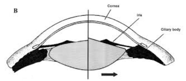
Orthokeratology utilizes custom-designed rigid gas-permeable contact lenses worn exclusively during sleep. These lenses temporarily reshape the anterior corneal epithelium through hydraulic pressure, flattening the central cornea to correct myopia while steepening the
mid-peripheral cornea. When the patient removes the lenses in the morning, their central vision remains clear throughout the day, while the ring of mid-peripheral corneal steepening creates continuous peripheral myopic defocus.
Defocus Incorporated Multiple Segments (DIMS) Spectacle Lenses
DIMS technology consists of a central clear zone for distance refractive correction, surrounded by a honeycomb-like matrix of hundreds of micro-segment “add” lenses (typically providing a relative positive power of +3.50 D). This structure simultaneously projects sharp central images onto the fovea while flooding the peripheral retina with a continuous planar field of myopic defocus.
Dual-Focus and Multifocal Soft Contact Lenses
Daily disposable soft contact lenses designed for myopia control feature alternating concentric rings of distance correction and treatment power (+2.00 D to +3.00 D add). Because the contact lens rests directly on the cornea and moves naturally with eye movements, peripheral myopic defocus is maintained consistently regardless of gaze direction.
6. Challenges and the Future of Myopia Theory
While relative myopic defocus remains the predominant theory guiding modern myopia management, ongoing research continuously refines our understanding of ocular growth regulation.
Individual Retinal Shape Variations: Retinal topography varies significantly between individual patients. An optical lens design that produces robust myopic defocus in one patient might produce less effective defocus in an eye with an unusually oblate or irregular posterior pole.
Visual Contrast Theory: Emerging scientific theories suggest that myopia control lenses may work not only through pure focal shifting, but also by reducing peripheral spatial contrast, which prevents hyperopic signal processing in retinal amacrine cells.
Combination Therapies: Modern pediatric eye care frequently combines optical RMD mechanisms (such as Ortho-K or DIMS lenses) with pharmacological agents like
low-concentration atropine (0.01%–0.05%), achieving synergistic control rates exceeding 70% by targeting both optical and biochemical growth pathways simultaneously.
Conclusion: A Paradigm Shift in Pediatric Eye Health
The discovery and validation of Relative Myopic Defocus transformed optometry and ophthalmology from a reactive field—simply prescribing stronger glasses every year—into a proactive clinical discipline capable of altering ocular development.
By understanding that the peripheral retina dictates the physical expansion of the eyeball, vision scientists turned an optical feedback loop into a powerful clinical tool. Today, leveraging relative myopic defocus through Ortho-K, advanced spectacle designs, and multifocal contact lenses gives children the ability to retain clear vision while protecting their long-term ocular health from the risks of high myopia.